
Fig 8.
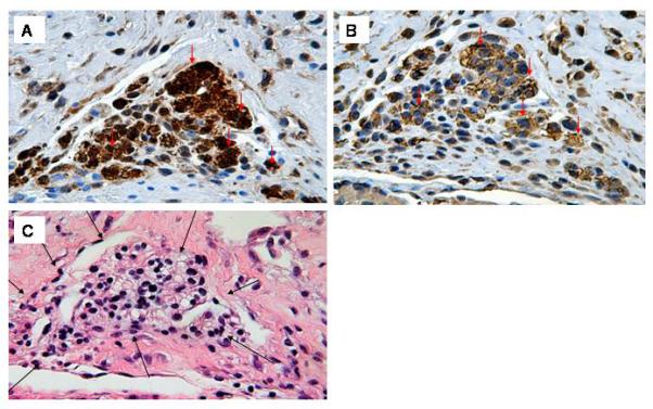
GFP and sarcomeric actin immune reactivity in hESC-derived cells 4 weeks following transplantation. A: Anti-GFP immune reactivity (HRP-conjugated secondary antibody, signal developed with diaminobenzidine reaction) (400×). B: Anti-sarcomeric actin immune reactivity on a serial section (HRP-conjugated secondary antibody, signal developed with diaminobenzidine reaction). Note that the preponderance of GFP-expressing hESC-derived cells also stain for sarcomeric actin (arrows) (400×). C: H&E staining of an adjacent section; note the absence of any lymphocytic infiltrate around the graft cells (black arrows) (400×).