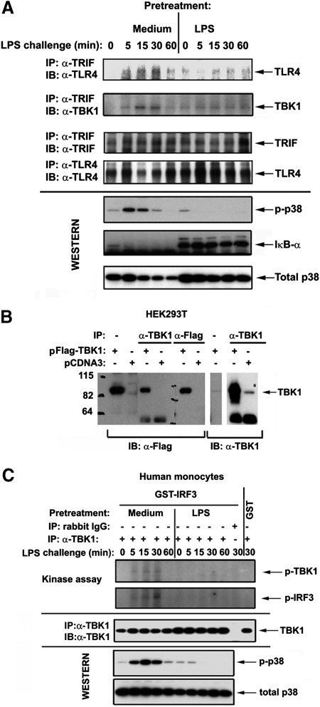
Figure 1.
Endotoxin tolerization dysregulates LPS-mediated TLR4-TRIF, TRIF-TBK1 associations and inhibits TBK-1 activation in human monocytes. After prior exposure to medium or 10 ng/ml LPS for 20 h, human monocytes were washed and treated with medium or challenged with 100 ng/ml LPS. (A) Whole cell lysates were prepared, immunoprecipitated with α-TLR4 (H80) or α-TRIF Abs, and immune complexes were analyzed by immunoblotting with the indicated antibodies. Shown are data of a representative (n=3) experiments. (B) HEK293T cells were transfected with either empty vector (pCDNA3) or pCDNA3-Flag-TBK1 and recovered for 24 h; whole lysates were prepared and immunoprecipitated with α-Flag or α-TBK1 Abs. Samples of whole cell lysates and Flag- or TBK1-containing immune complexes were fractionated by SDS-PAGE; proteins were transferred onto PVDF membrane and immunoblotted with α-Flag or α-TBK1 Abs. Numbers on the left indicate molecular mass standards (kDa). The results of a representative (n=2) experiments are shown. (C) Cells were lysed and whole cell lysates were immunoprecipitated with α-TBK1 Ab or isotype control IgG. Immunoprecipitates were subjected to in vitro kinase assay, using GST-IRF3 and GST recombinant proteins as substrates (top two panels) or to Western blot analyses with α-TBK1 Ab to control total expression of endogenous TBK1 proteins. (A and C): Expression levels of p-p38, IκB-α and total p38 were analyzed in whole cell lysates by immunoblotting with the respective Abs to control for LPS inducibility/endotoxin tolerization. Shown are data of a representative experiment (n=3).

