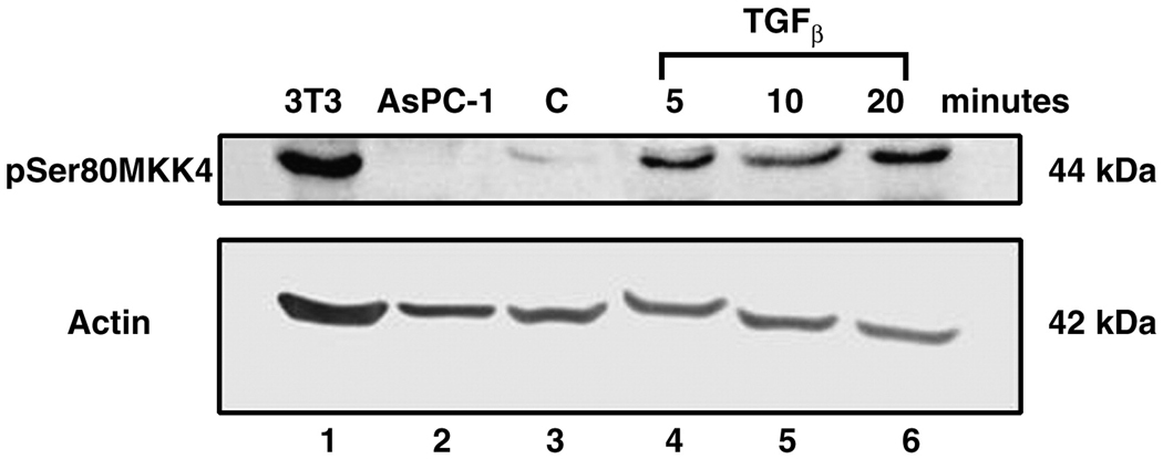
Figure 8. TGF-β induces MKK4 Phosphorylation in the SKOV-3 ovarian cancer cell line.
SKOV-3 cells were serum starved (control, lane 3) or treated with TGFβ (10 ng/ ml). Samples were harvested at the indicated time points (lanes 4–6). Duplicate Western blots were probed for either phosphoserine 80 MKK4 (top panel) or beta-actin (bottom panel). 3T3 cells are the MKK4 positive control (lane 1) and AsPc-1 cells are the negative control (lane 2). A 5.4 fold increase in serine 80 MKK4 signal was averaged across the 20 minute time course and normalized to the beta-actin signal. Beta-actin serves as a protein loading control (bottom panel).

