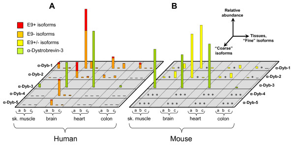
Figure 3.
Expression of α-dystrobrevin isoforms in human and mouse tissues. The relative expression of the discernable coarse and fine isoforms in A) human and B) mouse heart, brain, skeletal muscle and colon, as determined by quantitative RT-PCR. As indicated, horizontal axes show the tissues tested and the coarse and fine isoforms, while the vertical axis shows the relative expression level in arbitrary units. For human tissues, presence and absence of exon 9 (+ and -) was also assessed; these are shown as differently coloured portions of the bars. Asterisks in mouse indicate isoforms which while present in other mammals, are non-existent in murids. For display purposes, brain α-dystrobrevin-1a was used to normalise between species. Although α-dystrobrevin-3 is not directly comparable to the other reactions, heart α-dystrobrevin-3 has been normalised to brain α-dystrobrevin-1a, an approximate equivalence that is evident in published northern blots.

