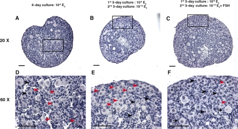
FIG. 2.
The histology of the primordial folliculogenesis in in vitro-cultured 17.5 dpc mouse fetal ovaries. A–C) Histology of 17.5-dpc mouse fetal ovaries cultured in vitro in 10−6 M E2 for 6 days (A); 10−6 M E2 for the first 3 days, then 10−10 M E2 for second 3 days (B); or 10−6 M E2 for the first 3 days and 10−10 M E2 plus 10 mIU/ml of FSH for the second 3 days (C). D–F) Higher magnification shows the structure of the unassembled oocytes, primordial follicles, and primary follicles in the ovaries cultured under the three conditions listed in A–C, respectively. Red arrowheads indicate unassembled oocytes (oocytes which were not surrounded completely by pregranulosa cells or by associated oocytes); black arrowheads indicate primordial follicles (one oocyte was surrounded by several flattened pregranulosa cells); and black arrows indicate primary follicles (one oocyte with expanded cytoplasm surrounded by a complete layer of cuboidal granulosa cells). Bar = 50 μm.

