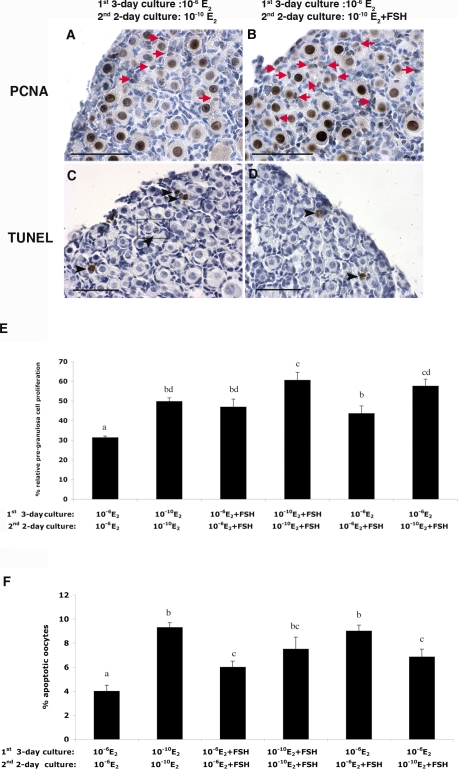
FIG. 4.
The proliferation of pregranulosa cells and oocyte apoptosis in 17.5-dpc mouse fetal ovaries after 5-day culture. PCNA (A) and TUNEL (C) staining of ovaries cultured in 10−6 M E2 for 3 days and then 10−10 M E2 for 2 days are shown, as are PCNA (B) and TUNEL (D) staining of ovaries cultured in 10−6 M E2 for 3 days and then 10−10 M E2 plus 10 mIU/ml of FSH for 2 days. Red arrows indicate PCNA-positive proliferating pregranulosa cells; black arrowheads indicate TUNEL-positive apoptotic oocytes. Framed areas indicate nuclear pyknosis in germline nests. E) Percentage of relative pregranulosa cell proliferation: PCNA-positive pregranulosa cells/total oocytes in one section of each ovary cultured under the indicated conditions (n = 12). F) Proportion of apoptotic oocytes: TUNEL-positive oocytes/total oocytes in one section of each ovary cultured under the indicated conditions (n = 12). Letters indicate statistically significant differences between treatment groups (P < 0.05). Bar = 50 μm.

