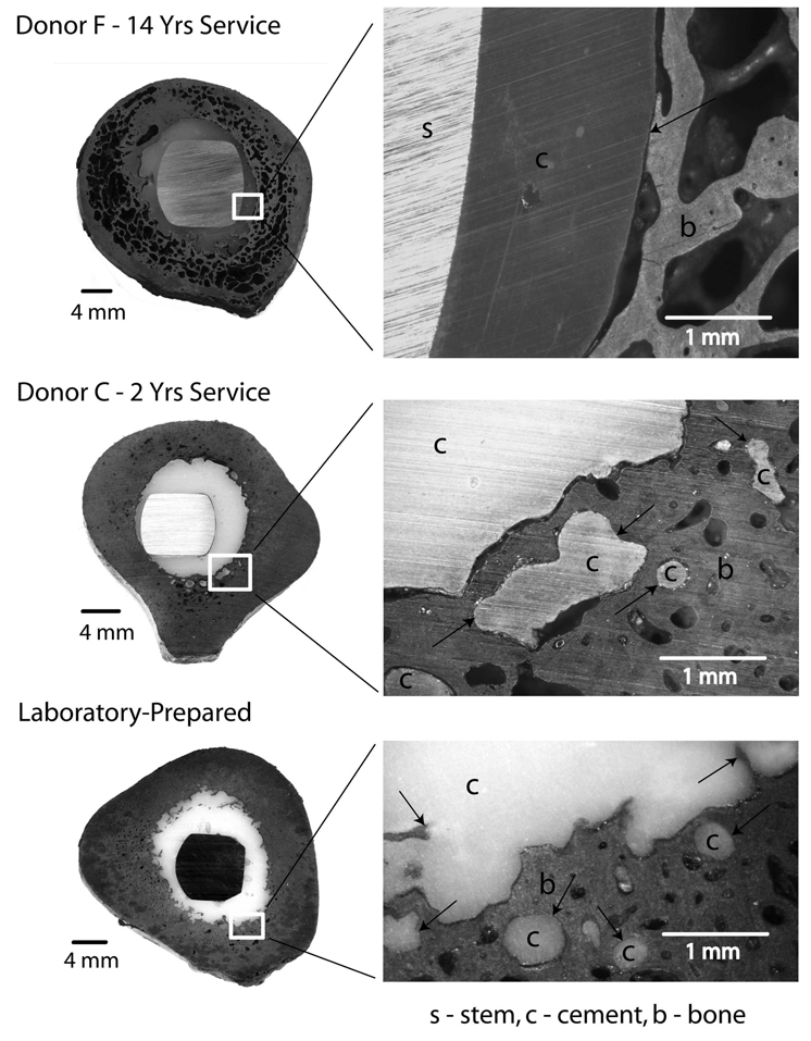
Figure 5.
Transverse section from donor bone F (70 mm distal to collar) illustrates extensive remodeling of the cortical bone and generation of a neocortex at the cement-bone interface. There are regions of cement-bone apposition along the interface. A section from donor bone C (120 mm distal to collar) shows much less cortical bone remodeling. There was cement flow into trabecular spaces and cortical bone lacunae and there were regions of apposition at the cement-bone interface. A section from a laboratory-prepared construct illustrates regions of cement-bone apposition and a very narrow gap between the cement and bone. Black arrows indicate areas of cement-bone apposition.

