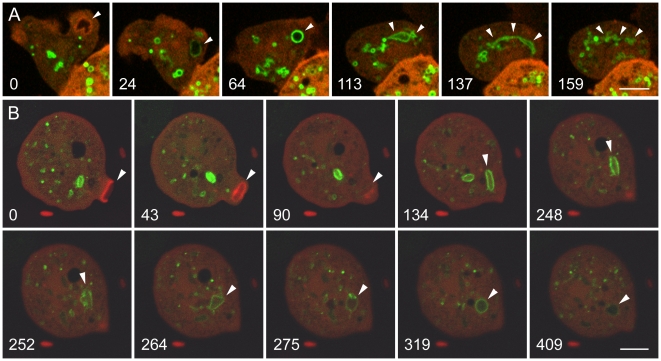
Figure 2. Labeling of early endosomes in Dictyostelium cells with GFP-2FYVE, a biosensor for PI(3)P.
The cells are expressing GFP-2FYVE and mRFP-LimEΔ. A, a macropinosome (marked by arrowheads in each panel). At 0 seconds, a nascent macropinosome is surrounded by actin filaments. After the actin filaments have disappeared, GFP-2FYVE begins to label the macropinosome, the strongest labeling being seen as the originally amorphous macropinosome assumes a spherical shape about 1 minute after uptake. During the next 2 to 3 minutes, the GFP-2FYVE label gradually fades as the macropinosome becomes increasingly elongated and fragmented. (See Movie S3.) B, phagosomes containing bacteria. The Dictyostelium cells were mixed with E. coli expressing a low level of a red cytoplasmic marker, and the uptake of two bacteria was tracked. In panel 0, the phagosome containing the first bacterium has already been internalized and has begun binding GFP-2FYVE. A second phagosome is beginning to form (arrowhead), surrounded by actin filaments (red); the completion and internalization of the phagosome is seen in the 43-second and 90-second panels. In subsequent panels, labeling by GFP-2FYVE reveals expansion and morphological changes in the phagosome, including tubular extensions in the 264-second panel. During this period, the GFP-2FYVE labeling fluctuates in intensity, being weaker at 275 seconds than at 319 seconds, and it has largely faded by 409 seconds. (See Movie S4.) A, Perkin-Elmer Ultra View; B, Zeiss LSM510 microscope. Bars, 5 µm.

