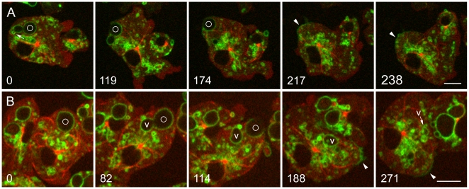
Figure 5. Retrieval of the V-ATPase following premature exocytosis.
A and B, Dictyostelium cells expressing GFP-α-tubulin and mRFP-LimEΔ were mixed with living S. cerevisiae 6 hours earlier. In both time series, a phagosome marked with a circle is exocytosed prematurely, prior to removal of the V-ATPase. A, at 0 seconds, the VatM-GFP-positive phagosome is close to the plasma membrane. A small vacuole brightly labeled with VatM-GFP (arrow) is in the process of budding off from the phagosome membrane. At 119 and 174 seconds, exocytosis is in progress. At 217 seconds, a patch of VatM-GFP is present in the plasma membrane at the site of exocytosis, and a microtubule is lying along the inner surface of the plasma membrane in that area. By 238 seconds, the VatM-GFP signal in plasma membrane is diminishing. The complete time series is shown in Movie S8. B, at 0 seconds, a VatM-GFP-positive phagosome is present near the plasma membrane. At 82 seconds, a large vacuole (V) enriched in VatM-GFP is separating from the phagosome. At 114 seconds, exocytosis of the phagosome is in progress, and at 188 seconds, it has been completed, leaving a bright patch of VatM-GFP in the plasma membrane (arrowhead). This label is much reduced by 271 seconds. (See Supplemental Figure S1 for quantitation.) Meanwhile, the VatM-GFP-positive vacuole (v) has moved about within the cell and undergone a series of morphological changes, including inward budding, which is evident in the 188 and 271-second panels. The complete time series is shown in Movie S9. Perkin-Elmer Ultra View microscope. Bars, 5 µm.

