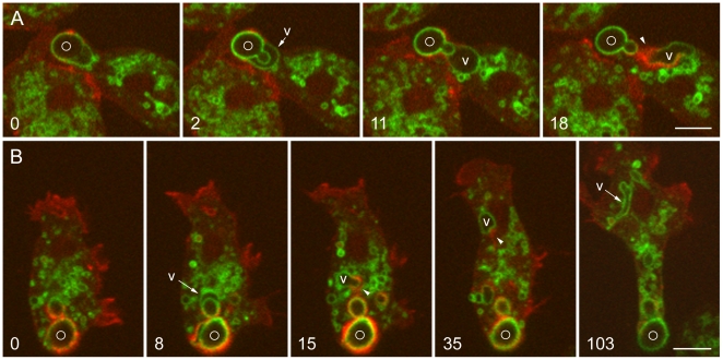
Figure 6. Acidic nature of prematurely exocytosed phagosomes and actin-powered vacuole movement.
A and B show Dictyostelium cells expressing VatM-GFP and DdmCherry-LimEΔ; the cells were mixed with living FITC-labeled S. cerevisiae 5 hours earlier. A, at 0 seconds, VatM-GFP is visible in the phagosome membrane, but the FITC-yeast inside can be seen only faintly. At 2 seconds, a VatM-GFP-positive vacuole (V) is beginning to form and the FITC-yeast has become bright; the yeast bud is now visible. At 11 seconds, the vacuole is separating from the phagosome membrane, and at 18 seconds, it is moving away. New actin assembly detected with DdmCherry-LimEΔ at the rear of the moving vacuole (arrowhead) appears to propel it. The complete time series is shown in Movie S10. B, at 0 seconds, a phagosome containing a budded yeast is faintly green but is more heavily labeled with the red probe for actin filaments. By 8 seconds, the green signal is stronger (appearing yellow where it overlaps the red) as the FITC-yeast has brightened, and a VatM-GFP-positive vacuole (V) has begun to separate from the phagosome. In the 15 and 35-second panels, actin filaments labeled with DdmCherry-LimEΔ can be seen at the rear of the moving vacuole (arrowheads), and by 103 seconds, the vacuole has become an irregular, elongated compartment. The complete time series is shown in Movie S11. Perkin-Elmer Ultra View microscope. Bars, 5 µm.

