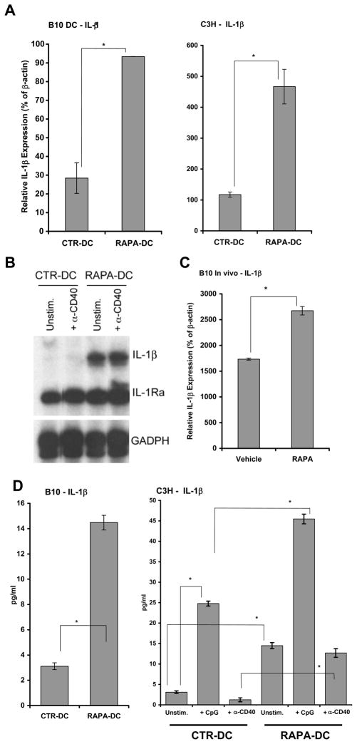
Figure 5. Exposure of DC to RAPA induces increased IL-1β expression.
(A) Significantly increased IL-1 β mRNA expression was observed for B10 (left panel) and C3H (right panel) CD11c+ RAPA-DC on d7 of culture by (A) qRT-PCR and (B) RPA. However, no corresponding increase was detected for IL-1R antagonist (IL-1Ra; B). Data are from single experiments representative of at least 3 separate experiments. (C) Significantly increased IL-1 β mRNA was also demonstrated for isolated CD11c+ splenic DC following 8d systemic administration of RAPA. Data are representative of at least 2 independent experiments. (D) Significantly increased IL-1 β secretion was also demonstrated for purified B10 (left panel) and C3H (right panel) RAPA-DC incubated for 18–22h (106 cells/ml) with or without CpG or α-CD40 mAb. All data are representative of at least 2 independent experiments.

