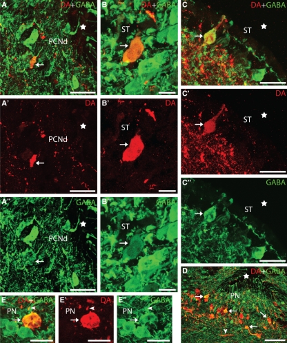
Fig. 3.
Transverse sections of the rostral diencephalon and telencephalon showing DA (red) and GABA (green) immunoreactivities. (A–A′′) Photomicrographs of a transverse section of the rostral diencephalon showing a double-labelled cell in the dorsal part of the postoptic commissure nucleus (arrows). (B–B′′) Photomicrographs of a transverse section of the striatum showing a double-labelled non-CSF-c cell of this region. (C–C′′) Photomicrographs of a transverse section of the striatum showing a double-labelled CSF-c cell. (D) Photomicrograph of a section of the preoptic region showing the high proportion of double-labelled (GABA/DA) dopaminergic cells. (E–E′′) Details of a double-labelled cell of the preoptic nucleus. Note the presence of a double-labelled terminal (arrowhead). Arrows indicate a double-labelled cell. Stars in figures indicate the impar telencephalic ventricle (A–C) and preoptic recess (D). Photomicrographs B–C′′ and E–E′′ are z-stack projections from eight confocal optical sections (0.5 μm thick). Dorsal is up in all figures. PCNd, dorsal postoptic commissure nucleus; PN, preoptic nucleus. For other abbreviations, see the legend to Fig. 1. Scale bars: 50 μm in A–A′′ and D; 12.5 μm in B–B′′; 15 μm in C–C′′; and 20 in E–E′′.

