
Figure 2. MHC molecule expression during IPCC development and following treatment with IFN-γ in vitro.
We generated IPCCs from ESF122 ES cells using the modified Blyszczuk protocol (see reference 2) and assessed the differentiation status of IPCCs in the penultimate (immature) and final week (mature) of the differentiation program. As expected, expression of the undifferentiated ES cell surface marker, SSEA-1, was negligible in both IPCC populations, verifying the fully differentiated status of IPCCs (data not shown)
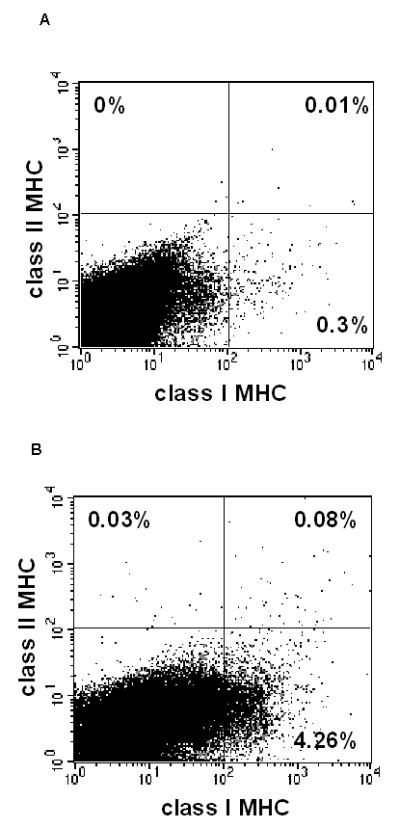
Immature IPCCs were stained for class I MHC and analysed by flow cytometry (A). This was repeated for mature IPCCs (B). MHC Class I expression increases to an average of approx. 4% cells in mature IPCCs, as assessed by flow cytometry. Gates for flow cytometry were set using unstained, isotype and no primary antibody controls. Data are representative of n=3 experiments.
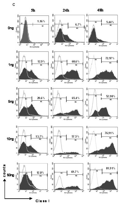
C: IPCCs generated using ESF 122 ES cells were assayed for expression of class I MHC molecules by flow cytometry. The IPCCs were stimulated in vitro with varying doses of IFN-γ over several time points to determine if the IFN-γ treatment would alter the IPCC expression of class I MHC. After the allotted time, the IPCCs were harvested and stained with the corresponding antibodies before acquiring the data on a flow cytometer. Gates were set using unstained, isotype (grey line) and no primary antibody controls. Representative flow cytometry dot plots from n=3 independent differentiation experiments are displayed.
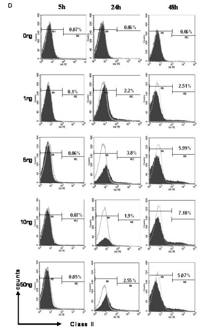
D: IPCCs generated using ESF 122 ES cells were assayed for expression of class II MHC molecules by flow cytometry. The IPCCs were stimulated in vitro with varying doses of IFN-γ over several time points to determine if the IFN-γ treatment would alter the IPCC expression of class II MHC. After the allotted time, the IPCCs were harvested and stained with the corresponding antibodies before acquisition on a flow cytometer. Gates were set using unstained, isotype (grey line) and no primary antibody controls. Representative flow cytometry dot plots from n=3 independent differentiation experiments. The experiments were repeated in another ES cell line, ESF 134, and similar results were obtained (data not shown).