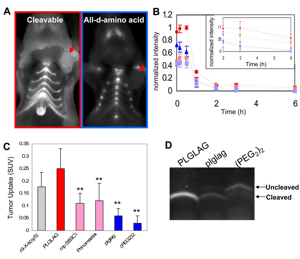
Figure 1. Uptake of free ACPP’s by mice harboring HT-1080 xenografts is protease dependent.
(A) Images of nude mice bearing HT-1080 xenografts six hours after injection with either cleavable ACPP or an all-D-amino acid control. Skin has been removed to expose the tumor (red arrows). (B) Shows a washout curve generated from taking regions over the tumor (red or blue) and thorax (pink or light blue, dashed lines) of animals receiving 10 nmol of either cleavable suc-e8-xPLGLAG-r9-c(Cy5) (n=3) (red diamonds or pink squares) or suc-e8-xplglag-r9-k(Cy5) (n=3) (blue triangles or light blue squares) respectively. Error bars represent standard deviations. (C) Tumor uptake of peptide in animals decreased when pre-injected with pharmacological inhibitors of MMPs (SB3CT or prinomastat). Uptake values for the r9-X-k(cy5) CPP probe as well as the all D-amino acid and 2 unit PEG linker control peptides are shown for reference. r9-x-k(Cy5) n= 3, PLGLAG n= 9, SB3CT n= 3, Prinomastat n= 3, plglag n= 8, mPeg n= 3. Error bars represent standard deviation. ** represents statistically significant difference from the cleavable ACPP using a two tailed t-test. (D) Polyacrylamide gel electrophoresis of tumor homogenates differentiates cleaved versus intact peptides recovered from tumors of IV injected mice.

