Figure 3.
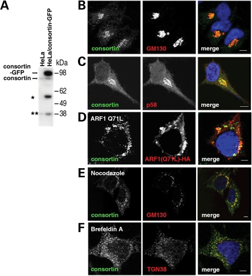
Subcellular localization of consortin in the Golgi apparatus. (A) Detection of consortin by an affinity-purified polyclonal antibody in lysates of untransfected HeLa cells (HeLa) or HeLa cells transiently transfected with a GFP-tagged human consortin construct (HeLa/consortin-GFP). Exogenously expressed consortin-GFP and endogenously expressed consortin are indicated by arrows. The lower molecular mass bands detected by the antibody (asterisks) may correspond to isoforms generated by translation started from a hypothetical internal ribosomal entry site or by post-translational processing. (B, C) Confocal microscopy images of HeLa cells stained for endogenous consortin (green) and the Golgi apparatus markers (red) GM130 (B) and p58 (C). (D) Modification of the distribution of consortin (green) in HeLa cells transiently transfected with the dominant negative Q71L mutant of human ARF1 (red). (E, F) Modification of the distribution of consortin in HeLa cells treated with Golgi-disrupting agents nocodazole (15 µm for 2 h) (E) and brefeldin A (5 µm for 45 min) (F); cells were stained for consortin (green) and for the Golgi markers (red) GM130 (E) or TGN48 (F). Bars, 5 µm.

