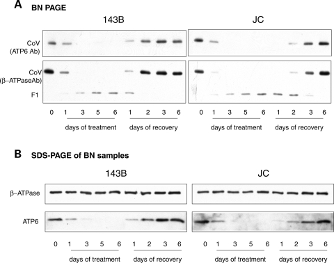
Figure 7.
Assembly kinetics of complex V. (A) BN-PAGE of complex V from 143B (left) and JC (right) cells grown and treated as in Figure 6. Samples were separated in parallel on two different gels, blotted and probed with antibodies against ATP6 subunit (top panels) and β-ATPase subunit (bottom panels). (B) Western blot of mitochondrial proteins solubilized as in (A), resolved by denaturing SDS–PAGE and detected with specific antibodies.

