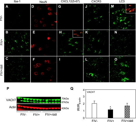
Figure 6.
Neuropathological changes associated with FIV infection and ddI treatment. Iba-1-immunoreactive microglia were detected in brains of mock-infected FIV− animals (A) but were more abundant and hypertrophied in FIV+ brains (B), and ddI treatment reversed this activation of microglia effect (C). NeuN-immunopositive neurons were detected in the parietal cortex of mock-infected controls (D), but were reduced in frequency in FIV+ animals (E), although ddI treatment appeared to protect neurons (F). CXCL12(5-67)-positive cells were not detected in mock-infected controls (G); in contrast CXCL12 immunoreactivity was observed in FIV-infected brains (H) but minimally detected in FIV-infected and ddI-treated animals (I). Inset shows colocalization of CXCL12(5-67) and NeuN in neurons. CXCR3 immunopositive cells resembling neurons were detected in mock-infected (J), FIV+ (K), and FIV+ animals receiving ddI (L). LC3-immunopositive cells colabeled with NeuN were readily found in mock-infected brains (M), but were reduced in number in FIV+ animals (N); ddI treatment appeared to reverse LC3 immunoreactivity in FIV infection (O). Synaptic protein VAChT was detected in brains of all groups (P). However, VAChT was reduced in FIV infection, but ddI treatment prevented the suppression of this synaptic protein (Q). Original view: ×630. *P < 0.05; Dunnet multiple comparison test.

