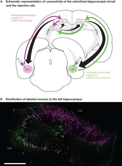
Figure 3.
Experimental design of dual viral tracing in the rat entorhinal-hippocampal circuit. (A) rHEP5.0-CVSG- β-gal and rHEP5.0-CVSG-EGFPx2 were injected to the left and right MEC, respectively. The viruses, transsynaptically transported from the bilateral MEC will go through CA1 and will first meet in the hippocampal CA3 region. (B) In field CA1, neurons are labeled only by the virus (rHEP5.0-CVSG- β-gal) injected into the ipsilateral MEC, whereas β-gal (magenta) and GFP (green) labels intermingle in CA3 region. Note that there are double-labeled neurons (white) in CA3. MEC, medial entorhinal cortex. Scale bar = 500 μm.

