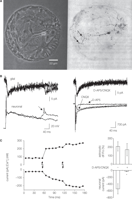
Figure 1.
Microisland cultures for studying neuronal–glial/astrocytic interactions. (A) Single neurons were grown on microislands consisting of glial cells, likely astrocytes (left, phase contrast). Cell permissive substrate, a mixture of collagen and poly-d-lysine, was aerosol micropatterned to form a spot onto an agarose coated glass coverslips. Hippocampal cell suspension was applied to dishes and cells adhered to permissive substrate forming microislands. Neurons were labeled (dark) with anti-synapsin I antibody (right, bright field); punctate staining (arrows) indicates putative autapses. Modified from Bekkers and Stevens (1991). (B) Neuron-to-glia signaling. Simultaneous electrical recordings obtained from a single hippocampal neuron (bottom) and astrocytes (top) residing on a microisland prepared as in (A). Glial cells (astrocytes) respond to synaptic transmission by an inward current (left), which represents an electrogenic activity of their plasma membrane glutamate transporters. Stimulation of a neuron to cause action potential evokes prolonged neuronal autaptic depolarization, which, in some cases, can drive an additional action potential (arrow) also causing a glial response. Ionotropic glutamate receptor antagonists (d-AP5 and CNQX) have no effect on astrocytic currents (right), while their use eliminated different components (slow and fast, respectively) of the autaptic currents in the voltage-clamped neuron. The asterisk indicates artifact due to application of a depolarizing step in the neuron. Modified from Mennerick and Zorumski (1994). (C) Astrocyte-to-neuron signaling. Neurons were plated onto pre-plated purified astrocytes forming microislands. Astrocytes occupying a microisland, loaded with the Ca2+ indicator and cage, were exposed to UV light (lighting bolts) to cause an increase in astrocytic [Ca2+]i. Left: Simultaneous electrical recordings from a single neuron, grown on top of astrocytes, indicate that the physiological increase in astrocytic [Ca2+]i is sufficient to cause glutamate-mediated SICs in neurons. Right: d-AP5/CNQX significantly (asterisk) attenuated the ability of photolytic Ca2+ elevations in astrocytes [shown as dF/Fo (%)] to cause neuronal SICs (right). Modified from Parpura and Haydon (2000).

