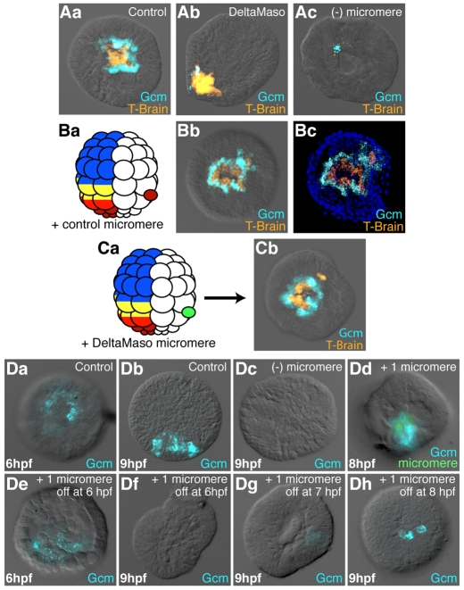
Fig. 5.
Regulation of Gcm expression by the Delta/Notch signal. (Aa-Ac) Effects of the loss of the Delta signal on Gcm expression. Co-staining by double-fluorescent in situ hybridization for T-brain (a micromere progeny marker, orange) and Gcm (blue) on control embryos (Aa), Delta-MASO-injected embryos (Ab) and micromereless embryos (Ac). (Ba,Bb,Ca,Cb) Role of Delta in the loss of Gcm expression in the Veg2U cells. (Ba,Ca) Schematics of the experimental design. A control micromere (Ba) or a DeltaMaso-injected micromere (Ca) was transplanted in between the veg1 and veg2 cell layers of a control 60-cell stage embryo. Gcm, blue; Tbr, orange. At pre-hatched blastula stage, 22 out of the 25 embryos (88%) transplanted with a control micromere showed ectopic expression of Gcm (Bb), whereas none of the Delta-MASO-injected micromeres induced ectopic Gcm expression (Cb). Bb and Cb are two-channel fluorescence and DIC images; Bc is a confocal projection of Bb with nuclear staining in blue. (Da-Dh) Duration of the Delta signal is crucial for the maintenance of Gcm expression. (Da,Db) Control embryos at 6 and 9 hours post-fertilization (hfp). (Dc) Micromereless embryos at 9 hpf. (Dd,De) Embryos in which all micromeres have been replaced by a single green dyed micromere at the 16-cell stage. In Dd, the transplanted micromere has been maintained up to fixation, in De it has been removed just before fixation. In Df, Dg and Dh, the transplanted micromere was eliminated at 6, 7 or 8 hpf, respectively, and the embryos were fixed at 9 hpf. (A-D) All embryos are in vegetal view, except for Ab, Db, Dd and De, which are in lateral view.

