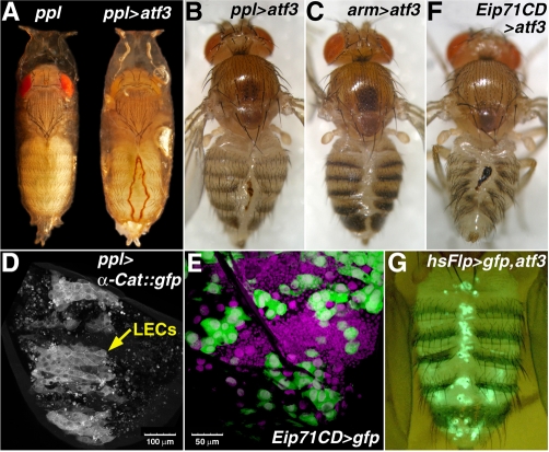
Fig. 4.
Misexpression of atf3 causes incomplete fusion of abdominal epidermis. (A) ppl>atf3 pharate adults usually die inside puparia with a dorsally open abdominal cleft in the adult cuticle. The scar is outlined with a brown line of necrotic tissue, and it is filled with the old pupal cuticle. Other body parts metamorphose normally and the unaffected regions of the abdomen produce adult cuticle with sensory bristles. (B) Few ppl>atf3 flies eclose, invariantly bearing the scar, often with signs of bleeding. (C) Effect of atf3 expressed under the ubiquitous (arm) driver. (D) Expression of UAS-α-Catenin::GFP in posterior abdominal segments of a normally developing pupa at 27 hours APF shows that the ppl-Gal4 driver is active in LECs but not in the surrounding histoblasts that occupy the areas around and between LECs at this time. The punctate GFP signals probably come from LECs that had already been extruded and phagocytosed. (E) The Eip71CD driver is also expressed in LECs (large GFP-positive cells) but not in histoblasts. DAPI staining (magenta) shows cell nuclei. (F) Abdominal lesion in a rarely eclosing Eip71CD>atf3 adult. (G) Flp-out induction of atf3 in the polyploid larval cells reproduces the abdominal closure phenotype (for more severe defects see Fig. S4 in the supplementary material). The LECs that express atf3 and GFP persist in the adult abdomen and mainly localize to the dorsal cleft. Scale bars: 100 μm in D; 50 μm in E.

