Fig. 1.
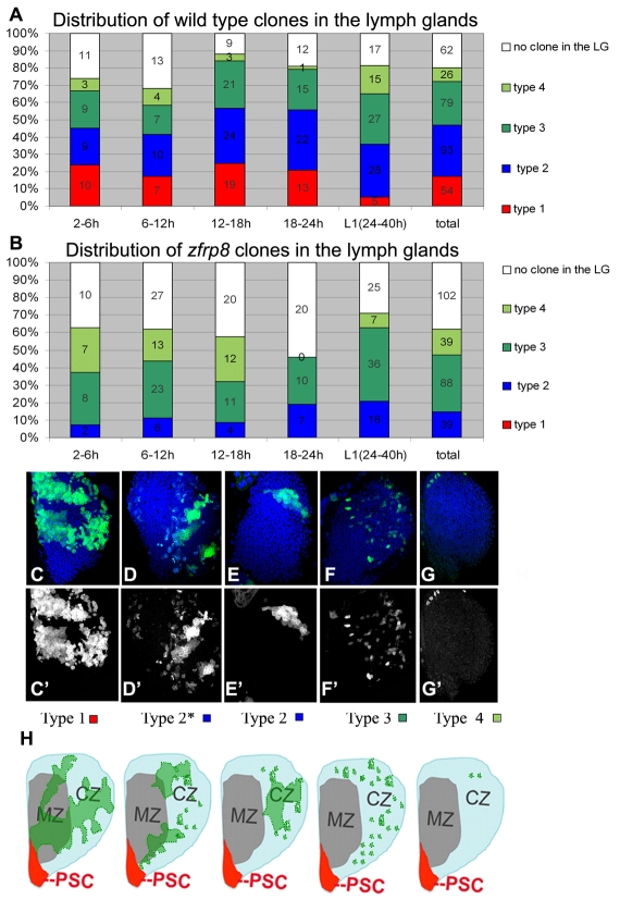
Four types of clones are identified in wild-type third instar larval lymph glands. (A) Wild-type clones. Proportion of MARCM clones generated at different time points after fertilization. Numbers of glands with each clone type and glands from mosaic animals with no clones are shown. Percentage (y axis) and numbers of each clone type (on column) are indicated. (B) Zfrp8 mutant clones. No type 1 clones are detected, and the percentage of type 2 clones is reduced. (C-G′) Confocal projections of mid-third instar lymph glands. (C,C′) Type 1 are cohesive clones, typically stretching from the medulla into the cortex and across all or some of the three to four cell layers (see intensity of GFP), occupying 10-30% of the lobe. (D-E′) Two examples of mixed population type 2 clones, located in different parts of the lymph gland, encompassing 4-8% of the lobe. (F,F′) Type 3 clones mainly consist of cells scattered in the cortex. (G,G′) Type 4 clones consist of one to eight cells, always in the cortex. (H) Schematics of third instar larval lymph gland primary lobes with different types of typical clones (green), based on multiple clones. (For more information, see Fig. 2.) PSC, posterior signaling center (red); MZ, medullary zone (gray); CZ, cortical zone (light blue).

