Fig. 4.
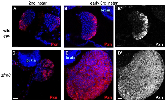
Hemocyte differentiation in lymph glands. Wild-type (A-B′) and Zfrp8 mutant (C-D′) lymph glands. Pxn (red) is expressed in the cortex of wild type (A) and Zfrp8 mutant (C) second instar lymph glands. The medulla of wild-type glands (A-B′) is defined by the absence of Pxn, whereas the cortex shows a sharp gradient of staining. Zfrp8 mutants show relatively normal Pxn staining in second instar lymph glands (C,C′) and rapid loss of medulla cells in early third instar glands (D,D′), where virtually all cells are Pxn-positive (note shallow gradient of staining attaining inner cortex levels of wild type). Scale bars: 20 μm.

