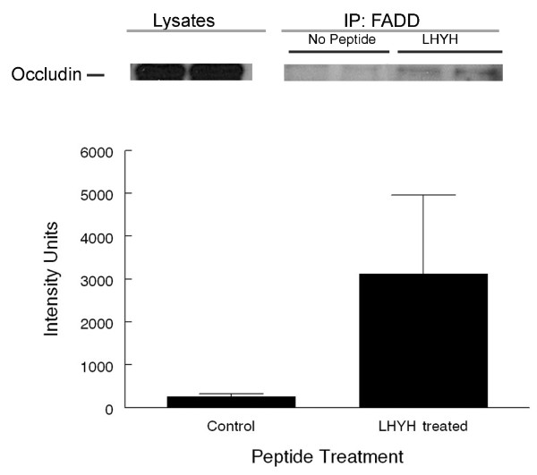
Figure 8.
Co-immunoprecipitation of occludin with FADD after treatment with LYHY. Eph4 cells were treated with 800 μM LHYH for 5 hrs or left untreated after medium change. Cells were lysed and proteins immunoprecipitated with a mouse anti-FADD antibody using protein A/G agarose beads and blotted with a rabbit anti-occludin antibody. A. Western blot for occludin. Lysates of cells that were immunoprecipitated with an anti-FADD antibody. IP-FADD: Two left lanes show IP of lysates from untreated EPH4 cultures and two right hand lanes show IP of lysates from LYHY treated EPH4 cultures. A very small proportion of the total cellular occludin was associated with FADD. B. Quantitation of occludin bands from 3 control and treated cultures. The intensity of stain was variable but higher in the immunoprecipitated fractions from LHYH treated cells (P < 0.1). Similar results were obtained from 2 comparable experiments.

