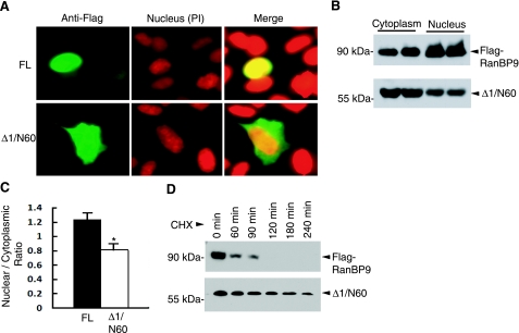
Figure 4.
RanBP9-Δ1/N60 is relatively more stable and cytoplasmic than RanBP9-FL. A) CHO-APP751 cells were transiently transfected with either RanBP9-FL or RanBP9-Δ1/N60 mutant. After 24 h, cells were stained with M2 antibody, followed by nuclear counterstaining with propidium iodide (PI). While RanBP9-FL was found in both the nucleus and cytoplasm, a significant proportion of cells showed exclusive localization of RanBP9-FL in the nucleus. B) HEK293FT cells were transiently transfected with either Flag-RanBP9-FL or Flag-RanBP9-Δ1/N60, and nuclear and cytoplasmic extracts were analyzed by immunoblotting with the anti-Flag antibody (M2). C) Quantitative measurement of intensity of bands in the nucleus and cytoplasm by Image J software revealed significantly less Δ1/N60 in the nucleus relative to cytoplasm compared with RanBP9-FL (means±se; n=4). *P < 0.01; Student’s t test. D) CHO-APP751 cells stably expressing Flag-RanBP9 or Flag-RanBP9-Δ1/N60 were treated with cycloheximide (100 μg/ml) for the indicated time points. Immunoblot analysis using anti-Flag antibody (M2) showed that the half-life of RanBP9-FL was <1 h, while that of half-life of Δ1/N60 was >3 h.

