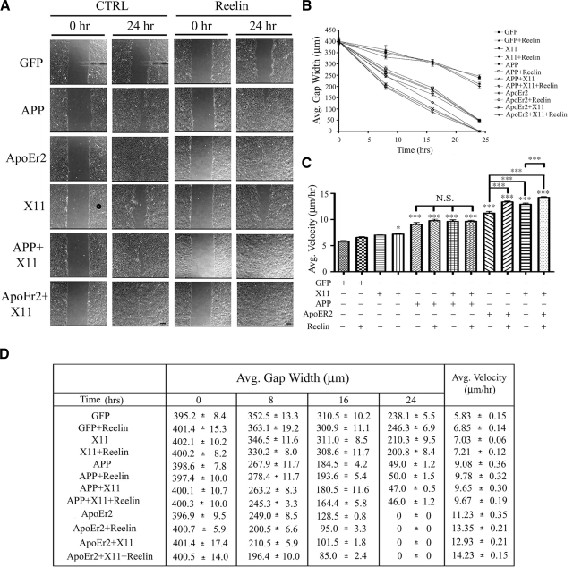
Figure 8.
Reelin increases the effects of X11α and ApoEr2 in a wound-healing assay. A) MCF 10A cells were transfected with constructs indicated at left and treated with control (left panels) or Reelin-conditioned medium (right panels) for 24 h. Representative images of the wound gap at 0 h (left) and 24 h (right) are shown. Scale bars = 75 μm. B) Average gap width was measured at 0, 8, 16, and 24 h following control or Reelin treatment. By 24 h, X11α or Reelin alone did not decrease gap width compared to GFP. X11α + Reelin slightly decreased gap width (16%; P<0.05); however, this decrease was significantly different from that of APP (79%), APP + Reelin (79%), APP + X11α (80%), APP + X11α + Reelin (81%), and all conditions, including ApoEr2 (100%) (P<0.001 for all). ApoEr2 further decreased gap width compared to APP alone (P<0.001), in the presence of X11α (P<0.01), with Reelin treatment (P<0.001), and in the presence of X11α + Reelin treatment (P<0.01). C) Average velocity of individual cells along the wound edge was measured over a 24-h period. Cotransfection of APP or ApoEr2 significantly increased average velocity of cells compared to control (56 and 93%) or X11α alone, and cotransfection of ApoEr2 with X11α resulted in a significant increase compared to ApoEr2 alone (122%). Reelin alone increased cell velocity in ApoEr2-transfected cells (19%) and also significantly increased cell velocity compared to control treatment of ApoEr2 + X11α-transfected cells (10%) (n=4). *P < 0.05, ***P < 0.001. D) Table of values showing quantification for B and C.

