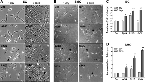
Figure 3.
Apoptosis induction by Aβ peptides in cerebral ECs and SMCs. ECs and SMCs were challenged with WT-Aβ40, E22Q, and L34V (50 μM) as described in Materials and Methods, and apoptosis was evaluated by phase-contrast microscopy and cell-death ELISA. A) Phase-contrast images of ECs both under control conditions and stimulated with Aβ peptides for 1 and 3 d. B) Phase-contrast images of SMCs both under control (Cnt) conditions and after challenge with Aβ peptides for 1 and 6 d. Arrowheads indicate cell shrinking; arrows indicate apoptotic bodies. C) Nucleosome formation by ECs challenged for 1 and 3 d with the respective amyloid molecules at 50 μM concentration, as evaluated by cell-death ELISA. D) Cell-death ELISA of SMCs treated for 1 and 6 d with 50 μM Aβ peptides. Apoptosis is expressed as fold of change compared with no-peptide controls. Data represent means ± sd of 3 independent experiments performed in triplicate. *P < 0.05, **P < 0.001 vs. respective no-peptide controls.

