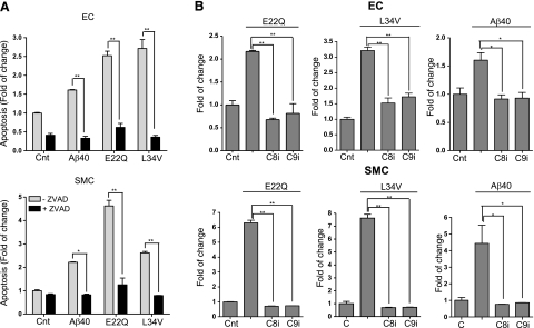
Figure 7.
Induction of caspase mediated apoptotic pathways by Aβ peptides. ECs and SMCs were treated with the different Aβ peptides at 50 μM concentration, in the presence and absence of either pan-caspase or specific caspase-8 or caspase-9 inhibitors, as indicated in Materials and Methods. Apoptosis induction in the presence and absence of the different pharmacological inhibitors was evaluated by cell-death ELISA, and results are expressed as fold of change compared with the no-peptide controls in absence of the inhibitors. A) Nucleosome formation in the presence or absence of the pan-caspase inhibitor Z-VAD (100 μM). Data represent 3 independent experiments. B) Nucleosome formation in the presence or absence of the specific caspase-8 (Z-IETD-FMK) or caspase-9 (Z-LEHD-FMK) inhibitors (both at 100 μM). Top panel: ECs; bottom panel: SMCs. Results represent means ± sd of 3 independent experiments performed in duplicate.*P < 0.05; **P < 0.001.

