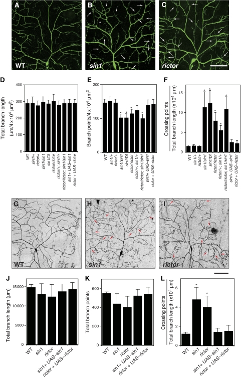
Figure 1.
sin1 and rictor function cell-autonomously in regulation of dendritic tiling in class IV neurons. (A–C) Live images of ddaC dendrites visualized by the pickpocket-EGFP (ppk-EGFP) reporter in wild-type (WT) (A), sin1PBac homozygote (B), rictorΔ2 homozygote (C). Anterior is left and dorsal is up. Arrows indicate crossing points of dendritic branches. Scale bar=50 μm. (D–F) Quantification of the total branch length (D), the branch number (E), and the crossing points (F) of WT and mutant ddaC dendrites. Error bars indicate mean±s.d. (WT, n=15; others, n=25), *P<0.01 (Student's t-test). Note that larvae heterozygous for sin1PBac over a deletion [Df(2R)BSC11] uncovering the sin1 gene show dendritic tiling defects identical to those of sin1 homozygotes. Genotypes: (A) yw; +/+; ppk-EGFP/ppk-EGFP, (B) yw; sin1PBac/sin1PBac; ppk-EGFP/ppk-EGFP, and (C) yw, rictor Δ2/yw, rictorΔ2; +/+; ppk-EGFP/ppk-EGFP. (G–I) MARCM clones of WT (G), sin1 (H), and rictor (J) are shown. Arrows indicate the crossing points of the dendrites. Scale bar=50 μm. (J–L) Quantification of the branch length (J), the branch points (K), and the crossing points (L) of MARCM clones. (WT, n=5; sin1, n=11; rictor, n=9) Clone genotypes: (G) hsFLP, elav-Gal4, UAS-mCD8-GFP/+; FRT42D, (H) hsFLP, elav-Gal4, UAS-mCD8-GFP/+; FRT42D, sin1PBac, AND (I) FRT19A, rictorΔ2; UAS-Gal4[109(2)80], UAS-mCD8GFP/hsFLP. Error bars indicate mean±s.d., *P<0.01 (Student's t-test).

