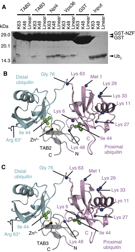
Figure 1.
Overall structures of the TAB2- and TAB3-NZFs in complex with K63-Ub2. (A) Pull-down assays using GST-fused TAB2-, TAB3-, Npl4- and Vps36-NZFs to assess their interaction with K63-, K48- or linear Ub2. The bound proteins were analysed by SDS–PAGE and stained with Coomassie Brilliant Blue. (B) The TAB2-NZF·K63-Ub2 complex. The NZF is coloured grey. The proximal and distal ubiquitin moieties are coloured pink and cyan, respectively. Ile 44 in each ubiquitin moiety is shown as green spheres. The Nɛ atoms of lysine residues and the nitrogen atom of the N-terminal Met in the proximal ubiquitin are shown as blue spheres. The K63R mutation in the distal ubiquitin is indicated as sticks. (C) The TAB3-NZF·K63-Ub2 complex. The drawing schemes are the same as in B.

