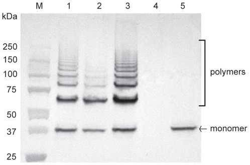
Figure 2. Western-blotting analysis of cell wall-anchored proteins of S. suis strain P1/7 and derived mutants with anti-Sfp1 antisera.
Mutanolysin-extracted surface proteins were resolved on Tris-HCl Ready gradient 4–15% SDS-PAGE gels (BioRad) and transferred onto nitrocellulose membranes. Sfp1 was detected using specific rabbit antisera and horseradish peroxidase-coupled, goat anti-rabbit secondary antibodies (Jackson ImmunoResearch, West Grove, PA). Five µg of protein were loaded per well. M: Molecular weight markers. Lane 1: WT strain P1/7. Lane 2: ΔsipF mutant. Lane 3: Δsfp2 mutant. Lane 4: Δsfp1 mutant. Lane 5: ΔsrtF mutant. See the results section for more details.

