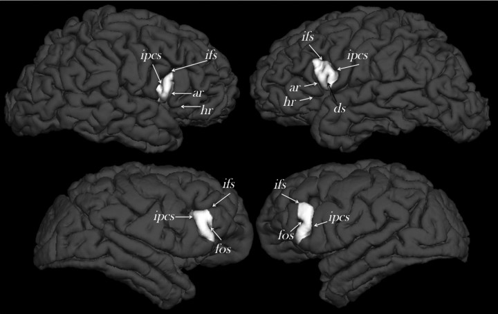
Figure 3.
External morphology of the right and left cerebral hemispheres of a randomly selected human (top) and chimpanzee (bottom) from the study sample indicating the location of the frontal operculum (white) and the defining sulcal contours (not to scale). Cortical reconstructions and labeling of the frontal operculum was performed using Freesurfer software (http://surfer.nmr.mgh.harvard.edu/). ar, Anterior ascending ramus of the Sylvian fissure; ds, diagonal sulcus; fos, fronto-orbital sulcus; ifs, inferior frontal sulcus; ipcs, inferior precentral sulcus.

