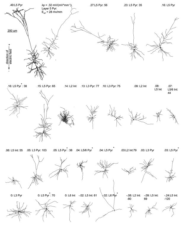
Figure 3.
Cortical neuron morphological reconstructions in order of electric field induced somatic polarization sensitivity. 3 items are listed for each cell, electric field induced somatic polarization length, λp (mm), an indicator of mV of polarization per unit electric field applied (mV/mm), layer, and cell type (pyramidal or interneuron); and if tested for that cell, electric field induced firing threshold. An asterisk next to the label for cell type denotes a neuron with a cut dendritic tree, that has still been included in all analyses.

