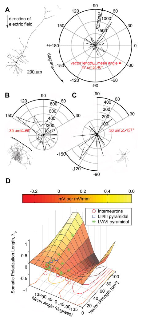
Figure 4.
Polar histogram coherence vector: Neuronal morphology predicts somatic polarization sensitivity to sub-threshold electric fields. A–C, Example tracings and corresponding volume-weighted polar histograms of (A) LV regular spiking pyramidal neuron with electric field The polarization length, λp (mm), an indicator of mV or polarization per unit electric field applied (mV/mm), = .32 mm, the polar histogram can be summarized by the variables: mean angle = 46° and vector length = 47 um3, representing the center of mass of the histogram; (B) LII fast spiking interneuron with polarization length = .14 mm, mean angle = 99° and vector length = 35 um3; and (C) LV fast spiking interneuron with polarization length = −.02 mm, mean angle = −127° and vector length = 30 um3. D, Summary plot of all neurons recorded and traced, with polar histogram coherence vectors as predictors of somatic polarization per electric field for each neuron. The colored plane is the statistically significant, best fit regression to the equation: polarization length = m*sine(mean angle)*vector length (p < .02, r2 = .41, n=30).

