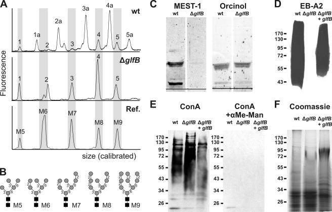
FIGURE 5.
The A. fumigatus ΔglfB mutant lacks Galf. A, electropherograms of fluorescently labeled N-glycans enzymatically released from secreted glycoproteins of A. fumigatus wild type (wt) and the ΔglfB mutant. Commercial oligosaccharides (Dextra Laboratories) served as reference (Ref.). The x axis was calibrated to the fragment sizes of the GeneScan-500 ROX standard (Applied Biosystems). B, schematic structures of reference oligosaccharides. Black squares, N-acetylglucosamine; gray circles, mannose. C, GIPCs extracted from A. fumigatus mycelium, separated by high performance thin layer chromatography, and stained with either the Galf(β1–6/β1–3)-specific antibody MEST-1 (left) or with orcinol/H2SO4 (right). The black line indicates the loading spot. D–F, water-soluble extracts of A. fumigatus mycelium separated by SDS-PAGE, transferred onto nitrocellulose membrane, and stained with the Galf-specific monoclonal antibody EB-A2 (D), mannose-specific lectin concanavalin A (ConA) in the presence or absence of 200 mm α-methyl-d-mannopyranoside (E), or Coomassie G-250 as a loading control (F).

