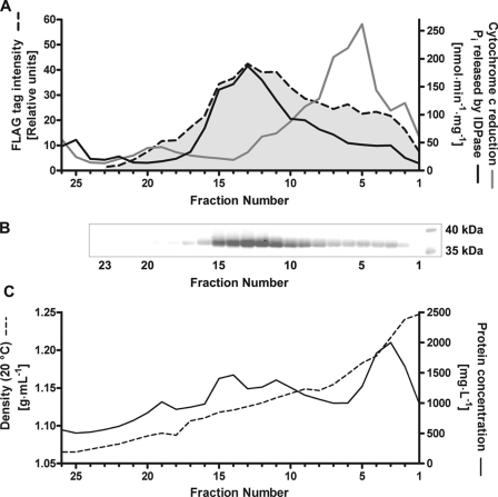
FIGURE 7.
Subcellular localization of GlfB in A. fumigatus. A. fumigatus FLAG-glfB microsomes were separated by isopycnic centrifugation on a multistep sucrose gradient. Fractions were analyzed by Western blot and immunostained with an anti-FLAG antibody (A and B) and compared with marker enzyme activities for endoplasmic reticulum (cytochrome c oxidoreductase, gray line) and Golgi (inosine diphosphatase (IDPase), black line) (A). Protein concentration and density were determined (C).

