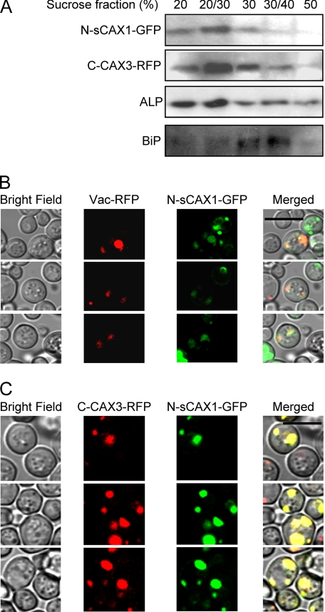
FIGURE 3.
Localization of CAX half proteins in yeast cells. Yeast cells expressing GFP-tagged sCAX1 N-terminal half protein (N-sCAX1-GFP) and/or RFP-tagged CAX3 C-terminal half protein (C-CAX3-RFP) were grown overnight in selection medium then diluted with water and observed using sucrose fractionation and confocal microscopy. A, discontinuous sucrose gradient fractionation of microsomes from yeast cells co-expressing N-sCAX1-GFP and C-CAX3-RFP. N-sCAX1-GFP and C-CAX3-RFP were co-fractionated in vacuolar membrane-enriched fractions (20% and 20/30% sucrose interfaces). Alkaline phosphatase (ALP) was used as a vacuolar membrane marker, and BiP was used as an endoplasmic reticulum marker. N-sCAX1-GFP was detected using a GFP antibody and C-CAX3-RFP was detected using an RFP antibody. B and C, co-localization of N-sCAX1-GFP with a prevacuole marker (Vac-RFP) (B) and co-localization of N-sCAX1-GFP and C-CAX3-RFP (C). The panels from left to right are: yeast cells in bright field, RFP image (Vac-RFP or C-CAX3-RFP), GFP image (N-sCAX1-GFP), and merged image. Bar, 10 μm.

