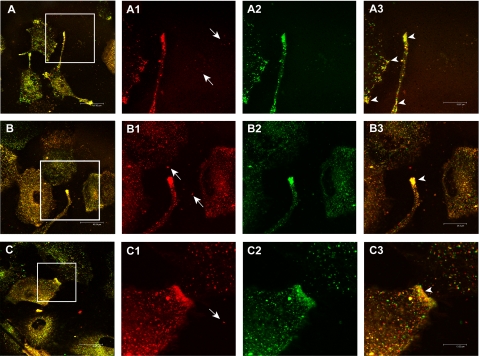
FIGURE 3.
In vivo exosome/melanoma lipids colocalization analysis through confocal laser-scanning microscopy. The panel shows three different fields (A, B, and C) of unfixed cell cultures in which co-cultivation of R18-labeled exosomes (red, magnifications in A1, B1, C1) and PKH67-labeled cells (green, magnifications in A2, B2, C2) is analyzed. In particular, arrows in A1, B1, and C1 images indicate free exosomes in cell culture medium not yet interacting with the cells. The yellow points (A, B, and C and arrows in magnification A3, B3, and C3) correspond to cell/exosome lipid mixing events, both at plasma membrane and intracellular levels. Bars: A, 48 μm; B, 46 μm; C, 42 μm; A3, 20 μm; B3, 24 μm; C3, 13 μm.

