FIGURE 5.
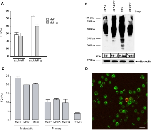
Role of microenvironmental pH in exosome fusion. A, R18-exoMel1 and R18-exoMel1ac were mixed with both acidic and buffered Mel1 cells (1 × 10⁶), and fusion efficiency was tested for 30 min. Points, means ± S.D. FD, fluorescence dequenching. B, NHS-biotin-buffered (pH 7.4) or acidic (pH 6.0) exosomes (25 μg) were incubated for 1 h with untreated or PPI-treated parental cells, then cells were analyzed by streptavidin blotting. Western blotting of nucleolin represents a control for cell protein equal loading. Numbers represent the whole lane densitometry analysis expressed in arbitrary units (a.u.). Points, means ± S.D. a p < 0.01 versus control (pH 6.0) (n = 3). C, R18-exoMel1 were added to metastatic melanoma (Mel1–3), primary melanoma (MelP1–3), and normal donor-derived PBMC, and fusion efficiency was tested for 30 min. Points, means ± S.D., (n = 3). D, R18-exoMel1 (red) were incubated for 3 h with PKH67-PBMC (green) and analyzed by confocal laser-scanning microscopy. The image clearly shows the absence of lipid colocalization areas. The rare event of colocalization observed in PBMC cells is likely due to the presence of monocytes, as recognizable from nuclear morphology. Bar, 20 μm.

