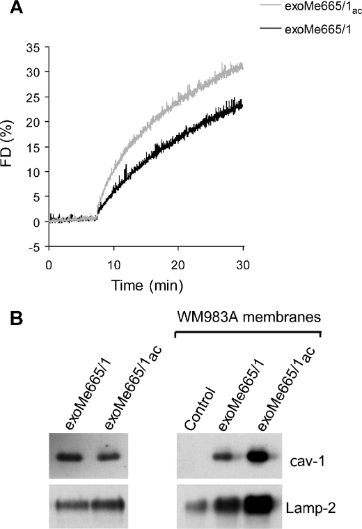
FIGURE 6.
Exosomes delivery of cav-1. A, R18-exoMe665/1ac and R18-exoMe665/1 (10 μg) were mixed with parental cells at the corresponding pH and fusion monitored. A representative fluorescence dequenching (FD) curve is shown. B, cav-1 and Lamp-2 immunoblotting on WM983A cell membranes after incubation with exoMe665/1 and exoMe665/1ac. Control represents WM983A membranes in the absence of exoMe665/1 incubation. A representative Western blot of two independent experiments is shown.

