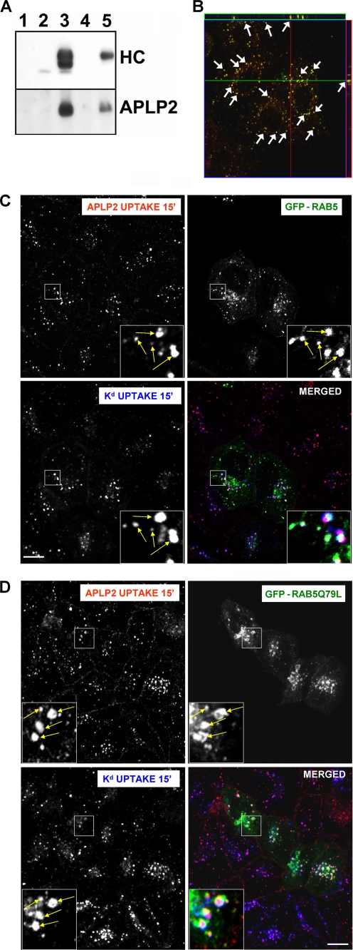
FIGURE 1.
APLP2 bound Kd at the cell surface and they internalized together into common Rab5+ early endosomes. A, Kd was demonstrated to bind APLP2 at the cell surface. Lane 1, HeLa-etKd cells were lysed and Protein A-Sepharose beads were added to the centrifuged lysate supernatant. Lanes 2 and 3, immunoprecipitations were performed with anti-Kd Ab 34-1-2 on HeLa and HeLa-etKd lysates. Lanes 4 and 5, HeLa and HeLa-etKd cells were incubated with Ab 34-1-2 on ice for 20 min, the cells were lysed and centrifuged, and Protein A-Sepharose beads were added to the supernatant. All samples were Western blotted with the 64-3-7 Ab to detect the etKd heavy chain (HC) or with anti-APLP2 Ab. B, Kd and APLP2 were identified as co-localized within vesicular compartments. Cell surface Kd and APLP2 molecules were labeled with 34-1-2 Ab and anti-APLP2 Ab, respectively, for 12 min at 37 °C to allow internalization of labeled proteins with the bound Abs, and any remaining Abs at the surface were removed. Cells were then fixed and stained with Alexa Fluor 568 goat anti-mouse Ab and Alexa Fluor 488 goat anti-rabbit Ab. Serial z-section images were acquired for HeLa-etKd cells, and the arrows point to common membrane structures on a representative z-section micrograph (obtained from six slices imaged at 0.4-μm intervals). Red, Kd; green, = APLP2; yellow, co-localized Kd and APLP2. C and D, cell surface Kd and APLP2 were internalized into the same endocytic vesicles. HeLa-etKd cells were transfected with either (C) GFP-Rab5 or (D) GFP-Rab5Q79L for 24 h, then pulsed with anti-Kd Ab 34-1-2 and anti-APLP2 Ab and incubated in complete medium for 15 min at 37 °C. Following the incubation, non-internalized Abs were removed. The cells were fixed, permeabilized, and stained with Alexa Fluor 568 goat anti-rabbit and Alexa Fluor 405 goat anti-mouse Abs. Red, APLP2; green, GFP-Rab5 or GFP-Rab5Q79L; blue, Kd; white, co-localized APLP2, Kd, and GFP-Rab5 or GFP-Rab5Q79L. Bar, 10 μm. The insets show a higher magnification of areas within the larger boxes, and the arrows point to representative endosomes with co-localized APLP2 and Kd.

