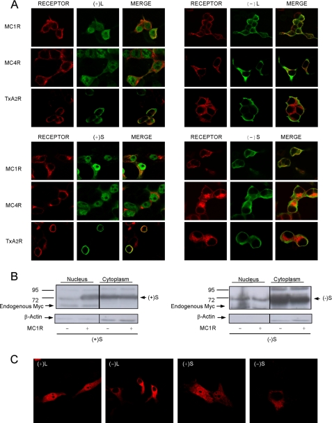
FIGURE 8.
Nuclear targeting of (+)-MGRN1 isoforms by MC1R and MC4R. A, HEK293T cells were transfected with MC1R, MC4R, or TxA2R and with Myc epitope-labeled MGRN1 isoforms, as indicated. The distribution of the proteins was visualized by confocal microscopy. Receptors are shown in red (left panel of each group). Middle panels, Myc-MGRN1s shown in green. The right columns in each group show the overlays. B, effect of MC1R on the presence of S isoforms in nuclear extracts. HEK cells transfected with Myc-labeled (+)S or (−)S MGRN1, alone or with MC1R, were fractionated into cytosolic and nuclear fractions. The fractions were analyzed for MGRN1 by blotting with αMyc. Also, blots were stripped and stained for β-actin. C, nuclear accumulation of (+)-MGRN1s in human melanoma cells. HBL cells were transfected with HA-labeled MGRN1s, permeabilized, stained with αHA, and analyzed by confocal microscopy.

