FIGURE 4.
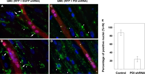
PDI is required for AChE localization on the cell surface. QMCs were co-transfected with red fluorescent protein (RFP) and either an EGFP shRNA as control (panels A and B) or a qPDI shRNA (panels C and D) to knock down PDI. Each group consisted of triplicate cultures, and the images were acquired from a separate culture under the same exposures. The white arrows point at cell surface AChE clusters on transfected cells in control group (panels A and B). The white asterisks highlight the remaining cell surface AChE on qPDI shRNA-transfected cells in the experimental groups (panels C and D). The white arrowheads point at cell surface AChE clusters on non-transfected cells in both control (panels A and B) and experimental group (panels C and D). Panel E, a bar graph shows the percentage of nuclei in multinucleated myotubes displaying AChE clusters on their adjacent cell surface in control and qPDI shRNA-transfected QMCs. All images were acquired using a 40× objective.

