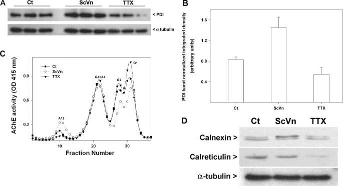
FIGURE 8.
PDI levels correlate positively with the levels of synaptic ColQ-AChE under different conditions of muscle activity. Sets of QMCs were treated for 48 h with ScVn or TTX. A, relative PDI content was determined by a Western blot of triplicate samples analyzed by SDS-PAGE and immunoblotted with anti-PDI and anti-α-tubulin antibodies. B, the integrated density of the PDI bands in A was determined and normalized versus the integrated density of the α-tubulin bands. C, the different AChE forms contained in the same volume of total cell extracts were fractionated by velocity sedimentation, and their enzymatic activity was assayed; solid circles, open circles, and solid triangles refer to control and ScVn- and TTX-treated groups, respectively. G1, G2, G4, A4, and A12 refer to AChE monomers, dimers, tetramers, collagen-tailed tetramers, and the ColQ-AChE form, respectively. D, relative calnexin and calreticulin content was determined by Western blot of pooled triplicate samples analyzed by SDS-PAGE and immunoblotted with anti-calnexin, anti-calreticulin, and anti-α-tubulin antibodies.

