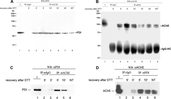
FIGURE 9.
PDI and AChE catalytic subunits form a complex during AChE folding. Lane 1 in each blot is a positive control consisting of an aliquot of the protein of interest. Lane 2 in each blot is the total cell extract from cells treated with DTT that were allowed to recover for 5 min and immunoprecipitated with the corresponding nonspecific IgG. Lanes 3–7 (A and B) and lanes 3–5 (C and D) are total cell extracts from cells treated with DTT that were allowed to recover from the treatment for 0, 5, 10, 15, or 30 min. Lanes 8 (A and B) and 6 (C and D) are total cell extracts from control cells not treated with DTT. A, QMC cell extracts were blotted directly with anti-PDI (lane 1) or after immunoprecipitation (IP) with anti-AChE (lane 3–8). WB, Western blot. B, parallel cell extracts were blotted directly with anti-AChE (lane 1) or after immunoprecipitation with anti-PDI (lane 3–8). C, HEK 293-hAChE cell extracts were blotted directly with anti-PDI (lane 1) or after immunoprecipitation with anti-AChE (lane 3–6). PDI levels are low in cells at 0 min after DTT treatment and in cells not DTT-treated. D, parallel cell extracts were blotted directly with anti-AChE (lane 1) or after immunoprecipitation with anti-PDI (lane 3–6). The higher apparent molecular size of the control protein in panel B, lane 1, is due to “smiling” of the gel. NT, not treated with DFP.

