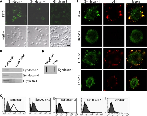
FIGURE 4.
Syndecan-1 binds to LG1 domain and in part to Ln2-P3 motif. A, expression of syndecan-1 and -4 and glypican-1 in PC12 cells. Cells were seeded on glass slide chambers for 2 days and immunostained with relevant antibodies (green). FITC, fluorescein isothiocyanate. B, immunodot blot analysis of the syndecan-1 and -4 and glypican-1 in PC12 cells. C, fluorescence-activated cell sorter analysis of PC12 cells assessing expression of syndecan isoforms and glypican-1 by using relevant antibodies (white area) compared with IgG-control (gray area). D, lysates were incubated with beads containing His6 alone or His6-rLG1, and the bound proteins were subjected to immunodot blotting and analyzed for syndecan-1 expression. Results are representative of three independent experiments. E, colocalization of rLG1 with syndecan-1. PC12 cells were seeded on glass slide chambers for 2 days, fixed, treated with heparin, scrambled peptide (Ln2-SP), Ln2-P3, or without the molecule (None) for 2 h at room temperature and further incubated with 25 μg/ml of rLG1 for 12 h at 4 °C. The cells were immunostained with anti-syndecan-1 antibody (green) and anti-His6 antibody (red). Scale bar, 20 μm.

