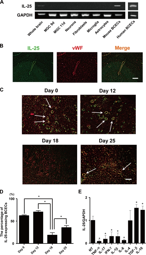
FIGURE 1.
IL-25 is expressed in BCECs. A, total RNA was extracted from the whole brain, primary cells, including mixed glial cells cultured for 9 and 11 days (MGC 9d and MGC 11d), neurons, astrocytes, microglia, mouse BCECs, and human BCEC lines. IL-25 and GAPDH mRNA expression was analyzed by RT-PCR. B, frozen brain sections were prepared from normal mice. After fixing with 4% paraformaldehyde, sections were stained against IL-25 (green) and vWF (red). Scale bar, 100 μm. C, frozen sections of the lumbar spinal cord were prepared from mice on the day of MOG/CFA immunization, 12 days (before the onset of EAE), 18 days (after the development of EAE), and 25 days (recovery phase of EAE) after immunization. Arrows show IL-25-expressing endothelial cells. Scale bar, 100 μm. D, results of C were quantified and graphed as the percentage of IL-25-expressing endothelial cells of total endothelial cells. E, MBEC4 cells were treated with or without TNF-α (50 ng/ml), IL-17 (50 ng/ml), IFN-γ (5 ng/ml), IL-1β (20 ng/ml), IL-6 (30 ng/ml), IL-4 (20 ng/ml), TGF-β (5 ng/ml), and IL-10 (20 ng/ml) for 24 h. Following RNA extraction, IL-25 mRNA expression was assessed by real time RT-PCR. The expression levels in untreated cells were set to 1. Data are represented as the means ± S.E. *, p < 0.05; n = 3. NT, not treated.

