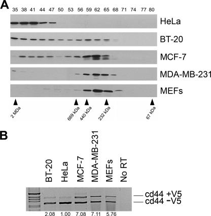
FIGURE 1.
Sam68 resides within a large and a small complex. The small complex correlates with the alternative splicing activity of Sam68. A, cellular extracts prepared from HeLa, BT-20, MCF-7, MDA-MB-231, and MEFs were fractionated over a Superose 6 column to separate complexes according to size. The molecular mass markers for the gel filtration column are shown below. The proteins from each fraction (numbered from 35 to 80 shown above) were separated by SDS-PAGE, and the presence of Sam68 was detected by immunoblotting with anti-Sam68 antibodies. The migration of Sam68 is shown. B, HeLa, BT-20, MCF-7, MDA-MB-231, and MEFs were transfected with a CD44 minigene, and the level of inclusion of the V5 exon was assessed by reverse transcriptase-PCR. The 1-kb ladder molecular weight markers (Invitrogen) are shown on the left. The ratio of inclusion (+V5)/exclusion (−V5) was quantified by densitometric scanning and normalized to 1.00 using HeLa cells.

