FIGURE 3.
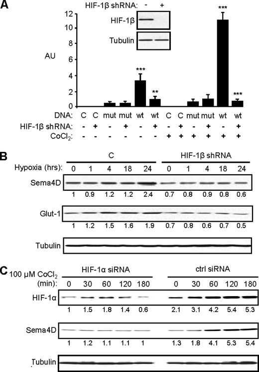
Sema4D protein levels increase in hypoxia in a HIF-1-dependent manner. A, HN12 exhibit reduced levels of HIF-1β protein when infected with lentivirus coding for shRNA against HIF-1β (inset). Cells expressing control shRNA (HIF-1β shRNA: −) or HIF-1β shRNA (HIF-1β shRNA: +) and either control DNA (DNA: C) or luciferase reporter plasmids containing the wild-type (DNA: wt) or mutated (DNA: mut) VEGF promoter were subjected to growth conditions with (CoCl2: +) or without (CoCl2: −) cobalt chloride for 2 h. A luciferase assay was performed with fluorescence (arbitrary units, AU) normalized by Renilla. Fluorescence was seen in cells expressing the wild-type luciferase construct and control shRNA lentivirus growing in CoCl2 but this was abolished by functional HIF-1β shRNA. Student's t tests were performed comparing cells expressing control or functional HIF-1β shRNA and wild-type reporter in normoxia or hypoxia, as shown, and p values calculated (**, p ≤ 0.01; ***, p ≤ 0.001). B, HIF-1β shRNA lentivirus-infected HN12 cells and control cells (C) were subjected to hypoxia (1% oxygen) for the times indicated and immunoblotted for Sema4D (upper panel) and Glut-1 (middle panel). Sema4D and Glut-1 protein levels increase in control cells growing in hypoxia, but these responses were lost in cells infected with HIF-1β shRNA-expressing lentivirus. C, increases in HIF-1α and Sema4D (upper and middle panel, respectively) observed in CoCl2-treated HN12 cells are lost in cells expressing HIF-1α siRNA oligos (HIF-1α siRNA) compared with controls (ctrl siRNA). Protein levels are quantified below each blot as a fold-increase relative to untreated controls (A) or untreated shRNA-expressing cells (B). Tubulin is used as a loading control in all immunoblots (lower panels).

