FIGURE 2.
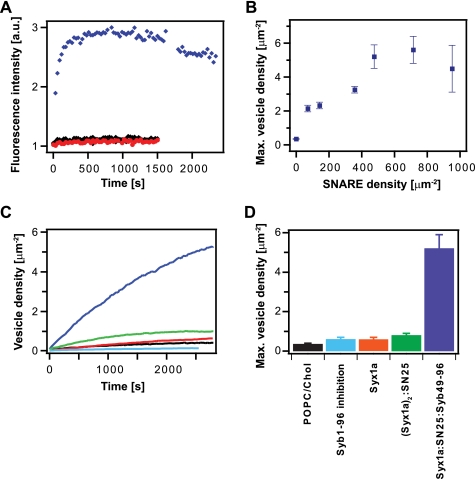
Specific binding of soluble synaptobrevin and docking of synaptobrevin vesicles to acceptor SNARE complex in planar-supported membranes measured by TIRF microscopy. A, binding of Alexa546-labeled Syb1–96 to acceptor SNARE complex (blue diamonds), Syx1a only (red circles) and protein-free bilayers (black circles). The proteins were reconstituted into planar POPC:cholesterol (4:1) membranes at a p/l of 1:1000. B, docking of Syb1–116 vesicles to acceptor SNARE complexes as a function of acceptor complex density in the bilayer. Docked vesicle numbers were calculated from the fitted final fluorescence intensities according to Equation 1 and then averaged from 3 to 13 independent experiments. Saturation was reached approximately at a p/l of 1:3000 corresponding to an acceptor density of 467 acceptor SNARE complexes/μm2 in the membrane. C, Syb1–116 vesicle docking curves measured on planar membranes containing acceptor SNARE complex (blue line), (Syx1a)2·SNAP-25 complex (green line), Syx1a only (red line), acceptor SNARE complex preincubated with Syb1–96 (light blue line), and on a protein-free lipid bilayer (black line). Proteins were reconstituted into planar membranes at p/l of 1:3000. D, mean final Syb1–116 vesicle densities after docking to planar membranes containing different target SNAREs at a p/l of 1:3000 as in C and averaged from 3 to 13 independent experiments.

